


Unveiling Facial Depths
Master Three-Dimensional Anatomy
With the aid of the iFace, you will gain a profound understanding of three-dimensional facial anatomy.
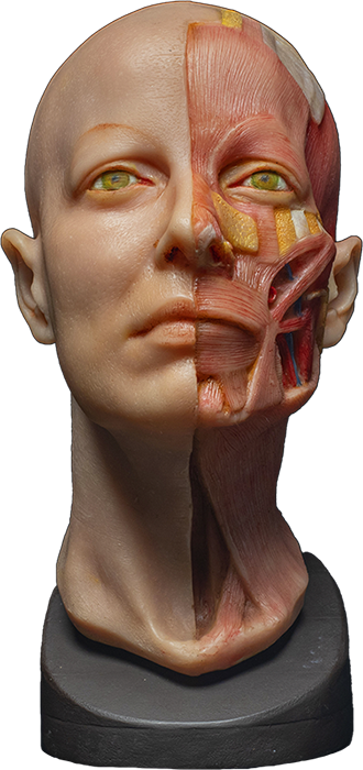
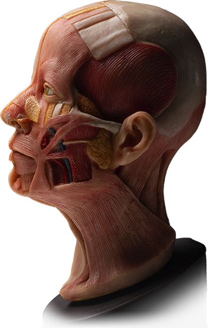
This model displays the face’s layers like no other, allowing you to comprehend the vital structures beneath the skin. Whether you work in surgical or non-surgical procedures, understanding these layers will guide your clinical practice.
This innovative model takes you on a journey through the layers of the face, starting with the scalp – SCALP representing the five layers: skin, superficial fat, galea aponeurotica, loose connective tissue, and periosteum. From there, explore other facial regions and their respective layers, gaining in-depth insights into the three-dimensional anatomy. This model also offers a unique advantage with its undissected side, allowing you to explore what lies beneath the surface in each specific region. With this newfound knowledge, you can confidently navigate facial procedures, ensuring both safety and aesthetic excellence.
3D Facial Layers Model Program: Mastering Three-Dimensional Anatomy of the Face
Curriculum Outline:
Module 1: Introduction
– Introduction to the Course
– Understanding the Importance of Three-Dimensional Anatomy
– Overview of the Windows E-Model and its Advantages
Module 2: The Layers of the Face
– Understanding the Scalp and Its Layers
– The Five Layers of the Hand-Bearing Area
1. Skin
2. Superficial Fatty Layer (Connective Tissue)
3. Aponeurosis and its Connection to Frontalis Muscle and Galea Aponeurotica
4. Loose Connective Tissue
5. Periosteum Covering the Entire Skull
– The Relationship Between the Deep Temporal Fascia and Temporalis Muscle
– The Frontalis Muscle and its Connection to Depressor Supercilii Muscle
– Continuous Connection of Frontalis Muscle with Orbicularis Oculi Muscle
– Identification of Preserved Nasalis Muscle and its Superficial Location
Module 3: Understanding the Mid-Face Region
– Exploring the Superficial Nasolabial Fat Compartment
– Layers and Anatomy of the Suborbicularis Oculi Fat (SOOF)
– The Fascia Separating the SOOF from the Bone
– Identification of Pre-Periosteal Fat within the Prezygomatic Space
– Boundaries of SOOF and Pre-Periosteal Fat
– Relationship between Zygomatic Minor Muscle, LLSAN, and Orbicularis Oris
– The Role of Zygomaticus Major and Risorius Muscle in Facial Expression
Module 4: The Infraorbital Region and Masseter Muscle
– Location of the Parotid Duct and Parotid Gland in the Preauricular Area
– Superficial Location of the Masseter Muscle and its Role in Jaw Movement
– Identification and Palpation of the Facial Vein
– Covering of Structures by the Platysma Muscle
– Connection of Buccinator Muscle to the Modiolus
– Understanding the Modiolus’ Connection with Orbicularis Oris Muscle
Module 5: Conclusion and Clinical Applications
– Summary of Key Concepts and Layers of the Face
– Application of Three-Dimensional Anatomy in Clinical Practice
– Guidelines for Safer and More Effective Clinical Outcomes
By completing the Windows E-Model Program: Mastering Three-Dimensional Anatomy of the Face course, you will not only acquire a unique educational tool but also gain a comprehensive understanding of facial anatomy and its layered structure. You can apply this knowledge to enhance your clinical practice and provide better care to your patients.
Unlock the potential of this remarkable model, and elevate your clinical practice with a profound grasp of facial anatomy. The future holds endless possibilities, and we look forward to witnessing your success with this extraordinary tool. Get ready to elevate your expertise and embark on a transformative journey into facial anatomy!